
Microarray
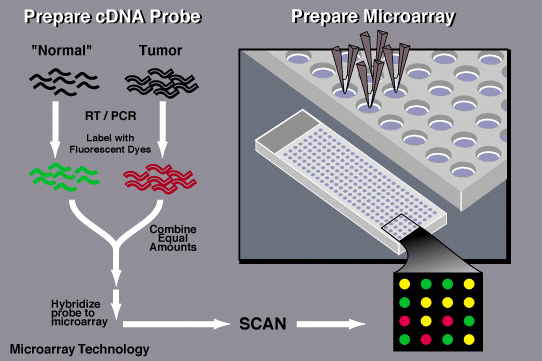
Extract the mRNA
mRNA present in cell is collected and placed into a test tube.

Create labelled mRNA
Reverse transcriptase enzyme (RT) generates a complementary cDNA to the mRNA. During the process, fluorescent nucleotides are attached to the cDNA. The tumor and the normal samples are labelled with different fluorescent dyes.


Transfer the labelled mRNA
Labelled mRNA is taken from the test tube and then spread onto DNA microarray slide.

mRNA binds to DNA
Hybridization occurred as the labelled cDNAs that represent mRNAs in the cell move around microarray and hybridize or bind to their synthetic complementary DNAs attached on the microarray slide, leaving its fluorescent tag.
Scanning the microarray
Red spot- the specific gene is more expressed in tumor tissue than in normal tissue
Green spot- gene is more expressed in the normal tissue
Yellow spot- the specific gene is equally expressed in normal and tumor tissues

How does microarray work?

Take tissue samples
Tissues are made of many cells. Each cell contains messenger RNA (mRNA). Tissue samples are taken from a patient.


This figure shows the steps of microarray.
A video of DNA Microarrays. Please enjoy it.